Liver Tumour Removal and surgical procedure
Liver Tumour Removal in Bangalore
Liver tumour removal, also known as partial hepatectomy, is a surgical procedure aimed at excising a portion of the liver affected by a tumour. This procedure is crucial for treating primary liver cancers as well as cancers that have metastasized to the liver from other areas, such as colorectal cancer.
Types of Liver Tumours:
- Hepatocellular carcinoma (HCC): The most common type of primary liver cancer.
- Metastatic liver cancer: Cancers that have spread from other parts of the body.
- Benign liver tumours: Such as hemangiomas, hepatic adenomas, and focal nodular hyperplasia.
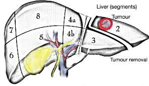
The Procedure for liver tumour removal:
Partial hepatectomy involves removing the tumour and surrounding liver tissue. The extent of liver resection depends on the size and location of the tumour. This procedure is performed by experienced (hepatobiliary) liver surgeon.
Body's Adaptability
Recovery from after liver surgery
Risks and Complications:
- Bleeding
- Infection
- Bile leakage
- Liver failure (rare)
Why Choose Us in Bangalore:
Our team of hepatobiliary ( bile ducts, gallbladder, liver ) surgeons is among the best in Bangalore, utilizing the latest technology and techniques for liver tumour removal. We offer comprehensive care from diagnosis to post-surgery recovery.
Deceased & Living Donor Liver transplantation
The surgical procedure that involves placing a liver graft either as a whole organ or a part is referred to as a liver transplant. Liver transplants have been categorised as
DDLT, deceased donor liver transplantation
This is also termed as Orthotopic transplant, which is the most commonly used procedure, wherein a whole liver is taken from a recently deceased donor and implanted in the patient. Meet our expert doctors for deceased donor liver transplantation in Bangalore.
LDLT, living donor liver transplantation:
A living-donor transplant is a surgical procedure to remove a portion of the liver from a living person and place it in another person whose liver is non-functional. The amazing property of the liver is that it can regenerate itself. This means that both the donor’s and recipient’s livers will regrow to their normal size within a few weeks after surgery. Meet our expert doctors for living donor liver transplantation in Bangalore.
There are three phases in the physiology of a transplant:
– Resection or ‘pre-anhepatic’ phase. Here liver dissection is carried out.
– Anhepatic phase. the period of time when no liver is in the circulation
– post-reperfusion phase). Also known as the Neohepatic phase, the stage at which the donor liver begins to function.
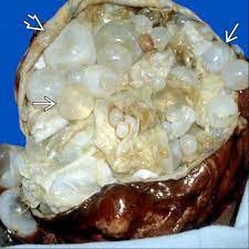
Hydatid Cyst:
The primary cause of hydatid cysts originates from tapeworms, a parasitic worm that lives in the guts of animals and humans. They enter the body in the form of larvae, and the eggs hatch into embryos that penetrate through the intestinal walls, get carried through the bloodstream to vital organs, and result in the formation of watery blisters. Identified as a hydatid cyst, with the liver being one of the potential organs to be affected. Hydatid disease is a probable fatal disease in the event of a lack of timely diagnosis and treatment. A heavily infested organ may fail or a cyst may rupture, resulting in a life-threatening allergic reaction.
The Symptoms of Hydatid Cyst are:
- Upset stomach.
- Unexplained weight loss.
- Abdominal swelling.
- Weakness and fatigue.
- Severe cough.
- Blood or the fluidic discharge while coughing.
The treatment for hydatid cysts is primarily a surgical procedure that is supplemented with anthelminthic medication to eliminate any leftover tapeworm eggs.
- Hydatid disease is a zoonotic and parasitic infection caused by by the larval form of the tapeworm Echinococcus granulosus. Most commonly it occurs due to accidental ingestion of tapeworm eggs excreted in the faeces of infected dogs.
- Hydatid disease is more prevalent in rural areas where there is a closer contact between people and dogs and various domestic animals which act as intermediate vectors.
- Hydatid disease can develop anywhere in the human body, the liver is the most frequently involved organ (50%–70%), folllowed by the lungs (20%–30%).
- It can asymptomatic for a long time,symptoms appear on increase in the volume of cyst or on development of complications.
- Right upper quadrant or epigastric pain,sensation of abdominal fullness, nausea, low-grade fever are the common symptoms
Patients with complicated cysts may present with
- Biliary communication- Classic triad of jaundice, biliary colic and urticaria
- Cyst rupture -Anaphylaxis secondary to the highly antigenic content of the cyst fluid or may be silent and present with multiple intraperitoneal cysts.
- Intrathoracic rupture- Dyspnoea and chest pain
- Secondary infection- Tender hepatomegaly, chills, and spiking temperatures.
Treatment for hydatid cyst varies from deroofing of hydatid cyst to resection of part of liver depends on the stage of the disease and associated complications caused by the disease under the cover of anthelminthic medication.
Liver Haemangioma:
Indications for Surgery:
- Progressive abdominal symptoms
- Spontaneous or traumatic rupture
- Rapidly enlarging lesions
- Kasabach–Merritt syndrome (Giant hemagioma and associated thrombocytopenia, anemia and coagulopathy)
- Unclear diagnosis
Various diagnostic Tests used to diagnose liver hematomas are:
- Ultrasound, an imaging method that uses high-frequency sound waves to produce images of the liver
- Computerised Tomography (CT) scanning, which combines a series of X-ray images taken from different angles around your body and uses computer processing to create cross-sectional images (slices) of the liver
- Magnetic resonance imaging (MRI) is a technique that uses a magnetic field and radio waves to create detailed images of the liver.
- Scintigraphy is a type of nuclear imaging that uses a radioactive tracer material to produce images of the liver.
Liver Trauma:
Most liver injuries occur from blunt trauma from road accidents, falls, bicycle crashes, violence, sports injuries that cause a blunt, forceful impact, or a penetrating injury that tears or cuts the liver.
Shunt Surgery for Portal Hypertension:
Extra Hepatic portal venous obstruction (EHPVO) and Non-Cirrhotic portal fibrosis (NCPF) causes Portal hypertension and Upper GI bleed. Surgery is indicated when bleed is refractory to medical management, Symptomatic Hypersplenism (Recurrent bleeds / infections), Symptomatic splenomegaly ( Pain, rupture, infarction, restriction of activities of daily living), Growth retardation, Portal biliopathy

Surgery involves Splenectomy (Removal of spleen) and formation of Proximal Spleno renal shunt. It selectively decompress gastroesophageal varices and maintain portal flow to the liver.